|
|
|
|
|
|
|
|
|
|
|
|
|
|
|
|
|
|
|
|
|

|
|

|
|
|
|
|
|
An Undergraduate Laboratory Project Exercise Integrating Actinomycetes Isolation from Soil with the Kirby Bauer Antibiotic Sensitivity Assay.
Outline
|
|
|
|
|
|
|
|
|
|
- Week 1. Usually the first laboratory meeting of the semester. Students are given an empty 35 mm film case and instructed to collect a surface soil sample from an area rich in organic matter.
- Week 2. The soil samples (approximately 5 grams) are placed in 100 mm empty sterile petri dishes and dried in a 37 C incubator for one week. This step reduces the "load" of non-spore forming bacteria and thus "enriches" the sample for desiccation resistant spore formers including Streptomyces, Bacillus, and Fungi.
- Week 3. This week's lab should be coordinated with a traditional laboratory exercise on serial dilution and spread plating. One gram of soil is serially diluted and 0.1 ml portions of 10-4, 10-5, and 10-6 dilutions are spread onto starch agar plates. Other culture media such as nutrient agar can be used but starch agar enhances the sporulation of the Streptomyces colonies. Incorporation of cyclohexamide into the culture medium reduces the growth of molds. The spread plates are incubated at 25 - 28 C.
- Week 4 or 5. Streptomyces colonies are often "late bloomers" and appear days after regular non-filamentous bacterial colonies. The use of a dissecting microscope aids in the identification of typical Streptomyces colonies ( 3 - 7 mm in diameter with depressions in the agar surrounding the colony). The colonies develop a fuzzy texture and often change color with the development of asexual conidiaspores. Zones of Inhibition of other bacteria are often (but not always) observed surrounding the colonies of antibiotic producing strains. Colonies are picked with an inoculating loop (moistened by dipping in a tube of sterile water) and streak plated onto fresh starch agar plates. By this time in the semester students are usually experienced at this technique. The plates are incubated at 28 C for two weeks to allow for the production of secondary metabolites including antibiotics. Students are encouraged to follow the appearance of the cultures during the incubation.
|
|
|
|
|
|
|
|
|
|
|
|
|
|
|
|
|
|
|
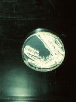
Figure 1. (left) A streak plate of Streptomyces isolate Ald13 on Starch Agar.
|
|
|
|
|
|
|
|
|
|
|
|
|
|
|
|
- Week 6. As soon as the Streptomyces colonies have sporulated on the streak plates, starch agar slide cultures can be prepared for microscopic examination of the filamentous mycelium of the isolates. This technique is usually done for the observation of molds and a suitable protocol is found in most introductory lab manuals. These slide cultures develop in less than a week. If 35 mm photomicroscopy equipment is available, students can take photographs of their isolates.
|
|
|
|
|
|
|
|
|
|
|
|
|
|
|
|
|
|
|
|
|
|
|
|
|
|
|
|
|
|
|
|
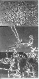
Figure 2. (left) Phase contrast photomicrographs of a slide culture of Streptomyces isolate Ald13.
|
|
|
|
|
|
|
|
|
|
|
|
|
|
|
|
|
|
- microcolony (400 x)
- branched hyphal filament (1000x) and
- branching filaments bearing asexual conidiospores (1000x).
|
|
|
|
|
|
|
|
|
|
|
|
|
|
|
|
|
|
|
|
|
|
|
|
|
|
|
|
|
|

|
|
|
|
|
Home Page | Streptomyces Project | Student Researchers
Recent Publications | Links to other Web Sites | Resume
Course Pages Virtual Office Hours
|
|
|
|
|
|

|
|
|
|
|
|
E-mail: stjohn@pop1.science.widener.edu
|
|
|
|
|
|